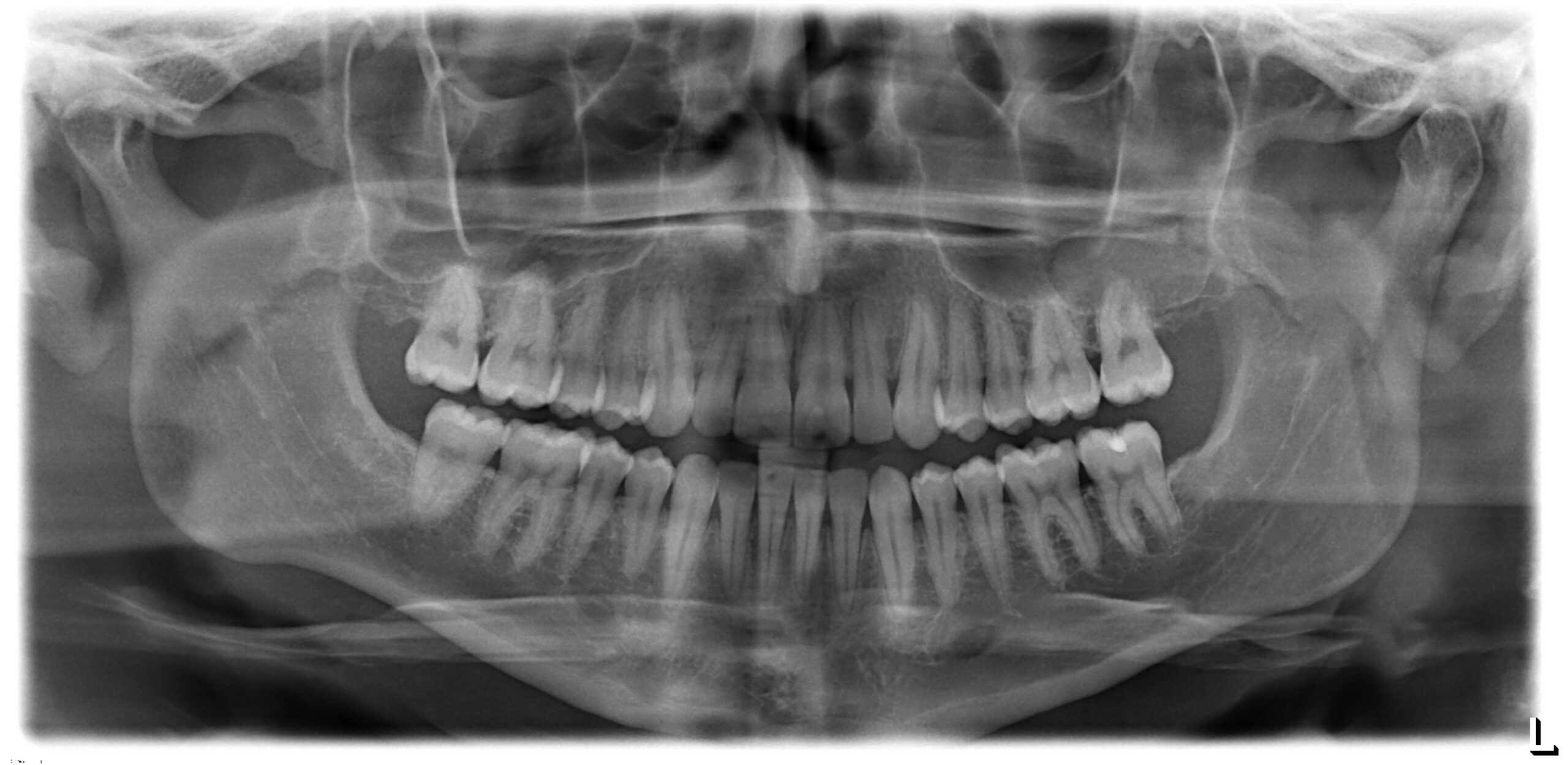
Are orthodontic X-rays safe? Yes. Modern digital orthodontic X-rays expose you to extremely low radiation levels, often less than a single day of natural background radiation from the environment. The American Dental Association, American Association of Orthodontists, and FDA all confirm that diagnostic dental imaging, when used appropriately, poses minimal risk to patients of all ages.
If you’ve ever wondered about the X-rays taken before braces or clear aligners, you’re not alone. Many patients and parents ask about radiation safety before starting treatment, and it’s a smart question to raise. Understanding what these images do, and how little radiation they actually involve, can put your mind at ease and help you make informed decisions for yourself or your child.

What Are Orthodontic X-Rays?
Orthodontic X-rays are diagnostic images that reveal what’s happening beneath your gums. They show teeth, roots, jawbone, and facial bone structure that can’t be seen during a visual exam alone. Think of them as a map your orthodontist uses to plan every move your teeth will make.
Your orthodontist uses several types of imaging:
- Panoramic X-rays capture your entire mouth in one wide image, showing all teeth, both jaws, and surrounding structures
- Cephalometric X-rays provide a side profile view of your skull, helping measure jaw relationships and growth patterns
- Cone beam CT (CBCT) scans create detailed 3D images for complex cases requiring precise measurements
- Bitewing X-rays may occasionally be used to check for decay or bone health around individual teeth before treatment begins
These images reveal critical information. Impacted teeth hiding beneath the gums. Root angles that affect how teeth can move. Jaw development patterns in growing patients. Without this view, creating an accurate personalized treatment plan would be guesswork, and small issues could turn into bigger ones down the road.
Most modern practices now use digital X-rays exclusively. Digital sensors capture images instantly and require far less radiation than older film methods. The difference is significant. Digital technology has made orthodontic imaging one of the lowest-radiation medical imaging options available today, while also giving providers sharper, more detailed pictures to work from.
How Orthodontic X-Rays Work and Radiation Exposure Levels
These images work by passing low-dose electromagnetic radiation through soft tissue. Denser structures like bone and teeth absorb more radiation, creating contrast in the image. The result is a detailed picture of structures you can’t see any other way.
Here’s what the numbers actually look like, based on approximate values reported by the ADA and FDA:
| X-Ray Type | Radiation Dose | Equivalent Natural Exposure |
|---|---|---|
| Digital panoramic | ~0.01 mSv | About 1 day of background radiation |
| Cephalometric | ~0.003-0.006 mSv | A few hours of background radiation |
| Full-mouth digital series | ~0.035 mSv | 3-4 days of background radiation |
| CBCT scan | 0.05-0.1 mSv | 1-2 weeks of background radiation |
For perspective, you absorb about 0.04 mSv of cosmic radiation on a cross-country flight. A chest CT scan delivers around 7 mSv. The average person receives roughly 3 mSv annually just from natural sources like soil, rocks, and cosmic rays.
Orthodontic imaging falls at the very bottom of the radiation scale.
Safety measures and modern protocols keep exposure low. Digital sensors require significantly less radiation than traditional film to produce a clear image, and clinicians use the smallest practical exposure needed for a diagnostic result. When shielding is used or requested, it should not interfere with image quality or require retakes.
Practitioners follow strict protocols set by the ADA and AAO. Every image serves a diagnostic purpose. Nothing is taken unnecessarily, and modern equipment is regularly calibrated to keep doses as low as reasonably possible.

Why Orthodontic X-Rays Are Necessary for Treatment
You might wonder if X-rays are truly necessary. Can’t an orthodontist just look at your teeth? The short answer: not completely, and skipping them could mean missing something important.
A visual exam shows only what’s visible above the gumline. Diagnostic imaging reveals everything else, from root angles to bone density to teeth that haven’t even come in yet.
What orthodontic X-rays uncover:
- Unerupted or impacted teeth that haven’t broken through the gums yet
- Missing teeth that may not develop at all
- Root length and angulation that determines how teeth can safely move
- Bone density that affects treatment timing and approach
- Jaw growth patterns critical for early treatment planning in children
- Cysts, tumors, or developmental abnormalities that require attention
- Previous trauma or hidden damage to roots that may not show symptoms
Without this information, your orthodontist would be working blind. Clear aligners, metal braces, and expanders all require precise measurements to work effectively. Imaging shows what’s really going on under the surface.
During treatment, progress X-rays may confirm that teeth are moving correctly. Final images verify root positioning before braces come off. According to the American Association of Orthodontists, diagnostic imaging is essential for safe, effective orthodontic care, and skipping it can lead to outcomes that don’t last or that cause complications later in life.
Early imaging in children is especially valuable. The AAO recommends an evaluation by age 7, partly because X-rays can spot developmental issues while the jaw is still growing and easier to guide. Learn more about age-based treatment on our orthodontics for all ages page.
Orthodontic X-Ray Radiation Compared to Everyday Exposure
Putting radiation numbers in context helps. Here’s how orthodontic X-rays compare to radiation you encounter in daily life, using approximate values from FDA and radiology references:
| Radiation Source | Approximate Dose |
|---|---|
| Digital panoramic X-ray | 0.01 mSv |
| Cephalometric X-ray | 0.005 mSv |
| Cross-country flight | 0.04 mSv |
| Annual background radiation (U.S. average) | 3.0 mSv |
| Chest X-ray | 0.1 mSv |
| Chest CT scan | 7.0 mSv |
| Abdominal CT scan | 10.0 mSv |
A panoramic image delivers roughly one-quarter the radiation of a single cross-country flight. You’d need to take hundreds of orthodontic X-rays to equal the radiation from one chest CT scan, which means the cumulative exposure from a full course of treatment is still extremely low.
The numbers speak for themselves, and the comparison really does drive home how small these doses are. Diagnostic imaging in orthodontics sits at the lowest end of medical radiation exposure. When weighed against the diagnostic value these images provide, the benefit clearly outweighs any theoretical risk.
How Often Are Orthodontic X-Rays Taken?
Most patients receive just a handful of X-rays during their entire course of orthodontic treatment. Imaging is typically taken at the beginning to plan treatment, occasionally during care if the orthodontist needs to check progress on a specific concern, and again at the end to confirm everything has moved into the right place. That’s really all most cases need.
Typical imaging schedule:
- Initial consultation: Diagnostic X-rays establish your baseline and guide treatment planning
- Mid-treatment (if needed): Progress images taken only if complications arise or the orthodontist needs to verify movement
- End of treatment: Final X-rays confirm proper root positioning before removing braces or completing aligner treatment
The frequency depends on your specific situation. Complex cases involving impacted teeth or jaw surgery may require more imaging. Straightforward cases often need only initial and final X-rays, which is great news for anyone worried about exposure.
Orthodontists follow the ALARA principle: As Low As Reasonably Achievable. This means taking only the images necessary for safe, effective treatment, and nothing more. A good orthodontist won’t order X-rays just for the sake of it. They’ll explain exactly which images your treatment requires and why before anything is taken, so you’re never left guessing about what’s happening or why it matters.
Who Should Take Extra Precautions With Orthodontic X-Rays?
While orthodontic X-rays are safe for the vast majority of patients, a few groups benefit from extra consideration: pregnant patients, young children, people with thyroid conditions, and anyone who has recently had multiple medical scans. Here’s what each group should know.
Pregnant patients: Tell your orthodontist right away if you’re pregnant or think you might be. Elective X-rays are typically postponed until after delivery. If imaging is medically necessary, careful technique and any appropriate protection are used to keep exposure as low as possible.
Children: Kids receive specially calibrated low-dose settings appropriate for their smaller size. Digital equipment and proper technique keep exposure as low as possible. The AAO confirms that orthodontic X-rays are safe for children when taken with appropriate protocols.
Patients with thyroid conditions: Tell your orthodontist about any thyroid concerns before imaging. Your provider can explain current safety protocols and whether additional precautions are appropriate for your situation.
Those with frequent medical imaging: If you’ve had recent CT scans, medical X-rays, or other imaging, let your orthodontist know. While orthodontic X-rays contribute minimally to cumulative exposure, your provider can factor this into treatment decisions.
Healthy adults and teens: Face negligible risk from routine orthodontic imaging. The radiation levels are simply too low to cause concern.
Always share your complete medical history with your orthodontist. Recent imaging, ongoing treatments, and health conditions all help the care team provide the safest possible experience from start to finish.

Frequently Asked Questions About Orthodontic X-Ray Safety
Can orthodontic X-rays cause cancer?
The risk is extremely low. A single panoramic X-ray exposes you to less radiation than one day of natural background exposure. According to the American Dental Association, no evidence links diagnostic dental X-rays to increased cancer risk when used appropriately. The benefit of accurate diagnosis far outweighs any theoretical concern.
Are X-rays safe for children?
Yes. Pediatric patients receive age-appropriate, calibrated doses using digital equipment designed to keep exposure low. Protective protocols and modern imaging standards add another layer of safety. The AAO recommends early orthodontic evaluation by age 7, and diagnostic imaging is part of that process when needed.
Can I refuse orthodontic X-rays?
You can, but it limits what your orthodontist can see and diagnose. Without imaging, impacted teeth, root issues, and bone abnormalities may go undetected. This can compromise treatment outcomes and even lead to complications. If you have concerns, talk them through with your orthodontist openly.
How are digital X-rays different from traditional film?
Digital X-rays use electronic sensors instead of film, requiring substantially less radiation to produce a clear image. Images appear instantly on a screen, can be enhanced for better visibility, and are stored electronically. There’s no chemical processing involved. Digital technology is a big step forward for safety.
Do clear aligners require X-rays?
Yes. Accurate treatment planning for clear aligners requires diagnostic imaging just like braces. These images reveal root positions, bone structure, and any hidden issues that affect how your teeth can move safely. Skipping this step would mean planning treatment without complete information.
Are X-rays safe during pregnancy?
Elective dental imaging is typically deferred until after delivery. If X-rays become medically necessary during pregnancy, careful technique and any appropriate protection are used to keep exposure to a minimum. Always inform your orthodontist if you’re pregnant or trying to conceive.
Have questions about X-rays, braces, or clear aligners? Schedule a free consult with Hamilton Orthodontics or start with a virtual consult from home.